Maxilla and Mandible Bone Density in Sample of Normal Full Dentition Yemeni Adults: Cone beam computed tomography (CBCT) retrospective study Cone beam computed tomography (CBCT) retrospective study
Main Article Content
Abstract
Background and Objectives: The internal structure of bones is described in terms of their quality or density, which reflects a number of biomechanical properties, such as strength and elastic modulus. Bone density refers to the concentration of minerals, particularly calcium and phosphorus, present in a given volume of bone tissue. It plays a vital role in determining bone strength, contributing approximately 60% of the overall structural integrity of bones. The study aims were to obtain baseline data on bone density of the maxilla and mandible in normal Yemeni individuals across various anatomical regions, genders, and age groups.
Materials and methods: retrospective analyzed study for CBCT images of 150 mandibular and 150 maxillary jaws from adults with full dentition in Sana’a City, Yemen. Scans were acquired using the PaxFlex3DP2 system and analyzed with Ez3Di and THEIA software.
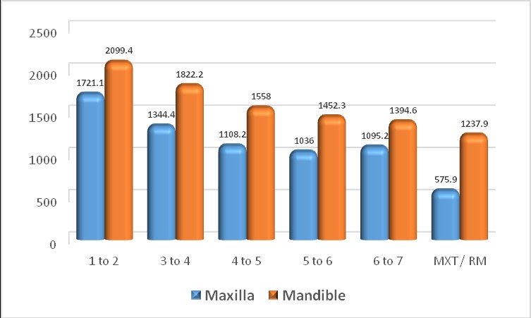
Results: The mean bone density values in maxillary and mandibular jaw for normal Yemeni people aged between (18-64y) ranged between (1036-1721 HU) for different maxillary areas and (1394.6-2099 HU) for different mandibular areas. The highest bone density was observed in both the mandible and maxilla between the central and lateral incisors, decreasing posteriorly in both jaws. Overall, no significant differences in bone density were found between males and females in most regions. However, in the maxilla, females showed significantly higher density between the first and second molars (P < 0.01). In the mandible, a significant difference was observed only between the central and lateral incisors, where females had higher density (P < 0.01). Age-related analysis revealed that the 31–45 age group had the highest bone density in both jaws (P < 0.01), with a gradual decline in older groups.
Conclusion: Bone density in Yemenis showed the highest values, one of the highest values compared to other nations. The lower jaw in Yemenis showed higher bone density than the upper jaw, with the highest values recorded in the anterior regions.
Downloads
Article Details

This work is licensed under a Creative Commons Attribution-NonCommercial-NoDerivatives 4.0 International License.

